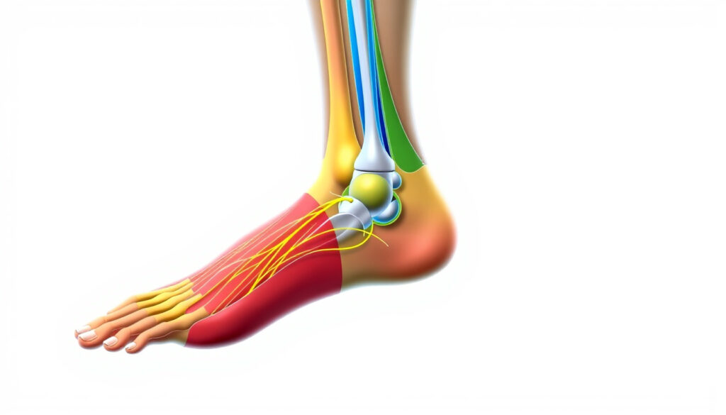
Lower Limb Dermatomes: Anatomy and Testing Guide
Most of us don’t give much thought to the skin on our legs — until a persistent ache, a patch of numbness, or a weird tingling sends us looking for answers. For clinicians and students alike, understanding lower limb dermatomes is the first step in mapping those sensations back to a specific nerve root.
Total human dermatomes: 30 ·
Lower limb dermatome range: L1–S2 (7 levels) ·
Most commonly impinged nerve roots: L5 and S1 ·
Standard dermatome map source: Clinically validated maps (e.g., Keegan & Garrett)
Quick snapshot
- Dermatomes are segmental skin areas innervated by a single spinal nerve root (Physiopedia (rehabilitation reference))
- Lower limb dermatomes span from L1 to S2 (Physiotutors (clinical education resource))
- Most common lumbar disc herniations affect L5 and S1 nerve roots (Physiotutors)
- Exact dermatome boundaries vary between individuals and between published map sources (Physiopedia)
- Overlap between adjacent dermatomes can make precise lesion localization imprecise (Physiopedia)
- Wallerian degeneration begins ~3 days after nerve injury (Physiotutors)
- Nerve regeneration starts at ~3 weeks, advancing ~1 mm/day (Physiotutors)
- Target reinnervation within 3 months if nerve gap ≤3 cm (Physiotutors)
- If sensory deficits are found, proceed to myotomal testing and reflex assessment
- MRI or electrodiagnostic studies may confirm nerve root compression
The table below summarizes the key numbers that anchor any discussion of lower limb dermatomes.
| Total spinal nerve pairs | 31 (8 cervical, 12 thoracic, 5 lumbar, 5 sacral, 1 coccygeal) |
|---|---|
| Lower limb dermatome count | 7 (L1 through S2) |
| Most common lumbar disc herniation | L5–S1 (affects S1 nerve root) |
| Nerve regeneration rate | Approximately 1 mm per day after Wallerian degeneration |
What are dermatomes of the lower limb?
Definition of a dermatome
- A dermatome is a strip of skin that receives sensory innervation from a single spinal nerve root (dorsal root) (Physiopedia (rehabilitation reference)).
Spinal nerve mapping overview
- 30 dermatomes exist: 8 cervical, 12 thoracic, 5 lumbar, 5 sacral (C1 has no dermatome) (Physiopedia).
- Lower limb dermatomes involve lumbar levels L1–L5 and sacral levels S1–S2 (Physiotutors (clinical education resource)).
Dermatome vs. cutaneous nerve distribution
- Dermatomes reflect segmental nerve root supply; cutaneous nerve territories reflect peripheral nerve branches. Overlap exists, but dermatome maps are the standard for localizing radiculopathy.
What are the dermatomes of the L4 L5 leg?
L4 dermatome distribution
- The L4 dermatome covers the medial leg and the medial foot, including the great toe (Physiotutors).
L5 dermatome distribution
- The L5 dermatome supplies the lateral leg and the dorsum of the foot, including the second through fourth toes (Physiotutors).
Overlap with adjacent dermatomes (L3, S1)
- Adjacent dermatomes overlap significantly, particularly at boundaries. This overlap can mask subtle deficits (Physiopedia).
The pattern in the table below is consistent: each nerve root controls a distinct strip of skin, and the reflex changes map directly to the same root.
| Nerve root | Key skin area | Associated reflex |
|---|---|---|
| L1 | Inguinal / groin region | None |
| L2 | Upper anterior thigh | None |
| L3 | Lower anterior thigh / knee | Patellar |
| L4 | Medial leg and medial foot (great toe) | Patellar |
| L5 | Lateral leg and dorsum of foot (toes 2–4) | None |
| S1 | Lateral foot and heel | Achilles |
| S2 | Posterior thigh and buttock | None |
The implication: knowing these landmarks lets you predict both the sensory territory and the reflex that will be affected when a specific root is compromised.
Where do you feel pain from L4 and L5?
Pain pattern for L4 radiculopathy
- L4 radiculopathy often radiates pain along the anterior thigh and down to the knee, sometimes with a reduced patellar reflex (Physiotutors).
Pain pattern for L5 radiculopathy
- L5 pain travels down the lateral leg to the dorsum of the foot, often accompanied by weakness in great toe extension (L5 myotome) (Physiotutors).
Differentiating L5 from S1 root involvement
- S1 pain follows a posterolateral leg route to the lateral foot and heel, and often blunts the Achilles reflex (Physiotutors).
L4 pain stays medial and high; L5 pain moves lateral and low; S1 pain lands on the heel. Reflex changes confirm the root.
How do you test for dermatomes in the lower leg?
Tools and materials
- Cotton wisp for light touch, sterile pin for sharp sensation, and optionally a tuning fork or cool/warm object (Clinical Physio (instructional video)).
Step-by-step testing procedure
- Position the patient supine with the limb exposed (Physiotutors).
- Start by demonstrating the sensation on the patient’s forehead as a reference (PMC / NIH (national medical library)).
- With the patient’s eyes closed, stroke each dermatome with a brush or cotton wisp, comparing the affected leg to the unaffected leg (Physiotutors).
- Test sharp sensation with a pin, applying even pressure over each key sensory point from L1 to S2 bilaterally (PMC / NIH).
- Ask the patient to rate sensation as equal, increased, decreased, or absent compared with the forehead (PMC / NIH).
Interpreting sensory findings
- Hypoesthesia in a single dermatome suggests a nerve root lesion; reduced pinprick sensation is more sensitive than light touch for single-level involvement (Physiopedia).
- Pooled sensitivity of sensory testing for lumbar disc herniation is about 35–40%, with specificity around 64% (2013 systematic review) (Physiotutors).
Sensory testing alone has limited rule‑in value (positive likelihood ratios 1.02–1.26). A negative test does not rule out radiculopathy.
How to remember the dermatomes of the leg?
Mnemonics for lower limb dermatomes
- A common mnemonic: “L1 groin, L2 upper thigh, L3 knee, L4 medial foot, L5 lateral foot, S1 heel.”
Clinical rules: the rule of 3
- In peripheral nerve injury: Wallerian degeneration at 3 days, regeneration starts at 3 weeks, target reinnervation possible within 3 months if the gap is ≤3 cm (Physiotutors).
Red flags in dermatomal pain
- Progressive motor weakness, saddle anesthesia (S2–S5), and bowel/bladder dysfunction require urgent imaging to rule out cauda equina syndrome.
What’s known and what’s uncertain
Confirmed facts
- Dermatomes are segmental skin areas innervated by a single spinal nerve root (Physiopedia)
- Lower limb dermatomes are well established in anatomical atlases
- Testing should compare both limbs and use both light touch and pinprick (Physiopedia)
What’s unclear
- Exact boundaries vary between individuals and published maps (Physiopedia)
- Overlap between adjacent dermatomes can make lesion localization imprecise
- Pooled sensitivity of sensory testing for disc herniation is modest (35-40%) (Physiotutors)
Key expert perspectives
“Pain examination begins by demonstrating the test on the forehead as a reference point. The examiner should prick the skin with even pressure over each dermatome from C2 to S5 bilaterally at key sensory points.”
— PMC / NIH (national medical library) standard procedure
“One recommended method is to stroke dermatome regions on both the affected and unaffected limb with a brush, such as the brush in a reflex hammer, and ask about side‑to‑side differences.”
— Physiotutors (clinical education resource)
For the clinician facing a patient with leg pain or numbness, the dermatome map is only half the story. The other half is knowing where the map blurs: individual variation and overlapping territories mean that a single sensory finding rarely pinpoints the exact root. The real diagnostic power lies in combining sensory, motor, and reflex data. For students and practitioners, the takeaway is clear: master the core landmarks (L4, L5, S1), use the rule of 3 to gauge recovery timelines, and never ignore red flags that point beyond a simple radiculopathy.
Frequently asked questions
What does dermatome pain feel like?
Patients often describe it as a burning, stabbing, or electric-shock sensation that follows a strip of skin. Numbness and tingling are also common.
What is the most commonly injured nerve in the leg?
The L5 nerve root is the most frequently compressed in the lumbar spine, often due to a disc herniation at L4/L5 or L5/S1 (Physiotutors).
What are the red flags of nerve pain?
Progressive weakness, saddle numbness, and loss of bladder or bowel control are red flags that require immediate further investigation.
What is the rule of 3 in a nerve injury?
Wallerian degeneration at 3 days, regeneration starts at 3 weeks, and target reinnervation within 3 months for gaps <3 cm (Physiotutors).
How do dermatomes differ from myotomes?
Dermatomes are sensory skin areas supplied by a single nerve root; myotomes are the group of muscles innervated by that same root. They are tested together via sensation and muscle strength.
More related posts
 Parker Posey: Queen of Indies, White Lotus Star Profile
 Alex Albon: Coma, Comeback, F1 Career & Net Worth 2025
 ANZ Banking Login – Step-by-Step Guide for AU and NZ Users
 Robin Gunningham: Is Banksy? Evidence and Anonymity Explained
 Russell Morris: Biography and 2025 Farewell Tour
 Jeffrey Sachs: Biography, Theories, and Controversies (2025)
 Black Dahlia: The Unsolved Mystery of Elizabeth Short
 Willow Tree Ireland: Native Species, Growth, Folklore & Downsides
